(TI = 3 s, TR = 5 s, TE = 15 ms, FA = 20 deg)
Here is what an inversion recovery image with this protocol would look like:
Source:Jupyter Notebook
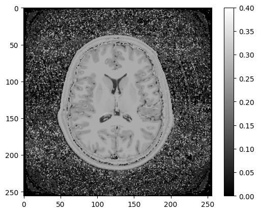
Figure 7.8:Inversion recovery image with TI = 3 s, TR = 5 s, TE = 15 ms, FA = 20 degrees
Do you think this is the correct answer? Can you see lesions?