The correct answer was A - (TI = 3 s, TR = 10 s, TE = 150 ms, FA = 90 deg). Here’s what this FLAIR image looks like:
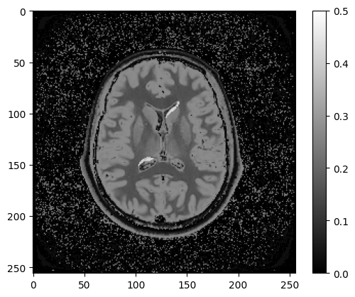
Figure 7.7:Inversion recovery image with TI = 3 s, TR = 10 s, TE = 150 ms, FA = 90 deg
Two periventricular lesions are clearly identifiable using this imaging protocol, which were hard to see on a regular T2w image Figure 7.2. The properly timed TI nulled the ventricular signal, whereas that long TR and TE provided sufficient T2 weighting to contrast against the nearby white matter, resulting in bright lesions, which are typically called hyperintese lesions.