 Quantitative MRI mOOC
B1 Mapping · Exercise 4-2
Registering qMRI measurements between each other: DAM example
A subject moved between their two DAM acquisitions: practice registering them together to get a good B1 map.
Exercise 4-2 · (a)
Register the double angle scans
A subject moved between their two DAM acquisitions: practice registering them together to get a good B1 map.
Note Recall the SCT lab from Lab 4
Layers top is drawn on top
Click and drag to move the second acquisition
Use the sliders to rotate or scale the brain
Mean error vs AFI
FA2 moved by
Your B1 map
Expected B1 (AFI)
How close to the expected B1 can you get? What’s the lowest mean error you can reach, and how?
Exercise 4-2 · (b)
The receive RF profile is multiplicative, and changes with position
The receive coils (orange) stay fixed in the scanner. Move and turn the head for scan 2: the two scans are then registered perfectly.
Scan 1 · FA1 = 60°
Scan 2 · FA2 = 120° · drag the head
Mean error vs AFI, perfectly registered
Head moved
Receive R2 / R1
B1 map
Notice
Every B1 technique that uses magnitude images depends on a ratio of measurements:
DAM: S2α / Sα
AFI: S2 / S1
Why are the receive coils not a problem if the subject doesn’t move, but a problem if they do?
The receive field R multiplies each image, at each piece of brain:
$$S_{\alpha} = R_1\,M_0\sin(\alpha B_1),\quad S_{2\alpha} = R_2\,M_0\sin(2\alpha B_1)\;\;\Rightarrow\;\;\frac{S_{2\alpha}}{2\,S_{\alpha}} = \frac{R_2}{R_1}\cos(\alpha B_1)$$No movement: R2 = R1, and it cancels. Movement: that piece of brain sat elsewhere in the coils’ field, R2 ≠ R1, and R2/R1 ends up in B1.
Exercise 4-2 · (c) · open question
How can we mitigate this in subjects who have difficulty staying still (e.g. pediatric, Parkinson’s)?
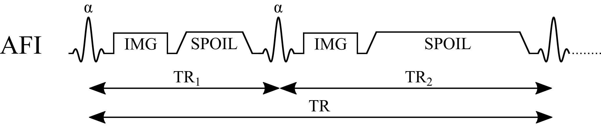
One solution: AFI
- A double angle takes two separate scans, minutes apart: the more the subject moves, the more R2/R1 ends up in B1, and registration can’t remove it.
- AFI acquires its two images in the same scan: each k-space line is read after TR1, then after TR2, a fraction of a second apart.
- So for each line the head is in the same place in both images: R2 = R1, and the ratio r = S2/S1 cancels the receive field. No motion between the images.