Introduction
Monoexponential T2 mapping
The simplest technique for performing T2-mapping involves the acquisition of multiple images using a single-echo sequence with different echo times (TE). This set of data can then be used to fit the T2 from the decaying signal curve Milford et al., 2015. However, this method is typically not employed in-vivo due to its prohibitively long acquisition times.
Multi-spin echo (MSE) sequences, based on the Carr-Purcell-Meiboom-Gill (CPMG) pulse train Carr & Purcell, 1954Meiboom & Gill, 1958, employ multiple 180-degree refocusing pulses to generate multiple echoes within a single acquisition (Figure 3.2) Fatemi et al., 2020Milford et al., 2015. This substantially reduces the scan time relative to acquiring multiple echoes using separate single-SE acquisitions, making MSE the sequence of choice in clinical settings.
The original Carr-Purcell method, which involves applying a 90-degree pulse followed by a series of 180-degree pulses along the same axis (e.g., x-axis), was designed to mitigate diffusion effects by preventing phase accumulation. However, this approach can lead to the accumulation of imperfections in the 180-degree pulses, resulting in a faster than expected loss of transverse magnetization (Mxy) and inaccurate T2 measurements Brown et al., 2014. The Meiboom-Gill improvement addresses this issue by applying the initial 90-degree pulse along one axis (e.g., x-axis) and the subsequent 180-degree pulses along a perpendicular axis (e.g., y-axis). This phase cycling distributes errors such that they average out over time, producing a more stable and accurate echo train Brown et al., 2014.
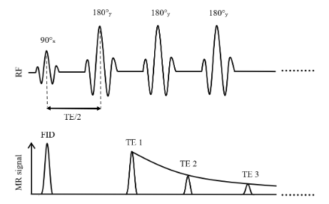
Figure 3.2:Simplified illustration of a multi-spin echo sequence for T2 mapping based on the Carr-Purcell-Meiboom-Gill method
To fit the data using the mono-exponential model, homogeneity within each voxel is assumed implicitly, implying a consistent T2 decay time across the tissue. This results in a single fitted T2 value for each voxel in the image. Although it is widely used, the mono-exponential model has been shown to be insufficient for proper estimation of tissue T2 relaxation times, given that the signal in a voxel often arises from multiple tissue components with different T2 values Graham et al., 1996. In a later section of this chapter, we will cover the multi-exponential method and discuss its use in applications such as myelin water fraction (MWF) imaging.
- Milford, D., Rosbach, N., Bendszus, M., & Heiland, S. (2015). Mono-exponential fitting in T2-relaxometry: Relevance of offset and first echo. PLoS One, 10(12), e0145255.
- Carr, H. Y., & Purcell, E. M. (1954). Effects of diffusion on free precession in nuclear magnetic resonance experiments. Phys. Rev., 94(3), 630–638.
- Meiboom, S., & Gill, D. (1958). Modified spin-echo method for measuring nuclear relaxation times. Rev. Sci. Instrum., 29(8), 688–691.
- Fatemi, Y., Danyali, H., Helfroush, M. S., & Amiri, H. (2020). Fast T2 mapping using multi-echo spin-echo MRI: A linear order approach. Magn. Reson. Med., 84(5), 2815–2830.
- Brown, R. W., C. Norman Cheng, Y., Haacke, E. M., Thompson, M. R., & Venkatesan, R. (2014). Magnetic resonance imaging: Physical principles and sequence design (R. W. Brown, Y.-C. N. Cheng, E. M. Haacke, M. R. Thompson, & R. Venkatesan, Eds.; 2nd ed.). Wiley-Blackwell.